The Shoulder
Shoulder on MRI
Magnetic resonance imaging (MRI) provides a detailed assessment of the soft tissues of the shoulder, including the rotator cuff, labrum, joint cartilage, among others. Defining what is “normal” on a shoulder MRI can be challenging, as many “abnormalities” noted in a radiology report may be changes that do not necessarily relate to a patient’s symptoms. This section aims to 1) orient the reader to the three different planes of a shoulder MRI and 2) highlight where to find key anatomic structures on MRI. These example images provide an efficient primer, however do not address the many nuances involved with interpretation of a shoulder MRI. Patients who undergo an MRI are provided an individualized and comprehensive explanation of their MRI findings during consultation with Dr. Brusalis.
An MRI provides a 3-dimensional view of the shoulder by providing 3 separate imaging planes.
The Coronal Plane is most similar to how we conceptualize the shoulder (i.e. viewing from the front) and resembles the AP X-Ray. Among the structures best visualized in this plane are the supraspinatus and infraspinatus tendons of the rotator cuff. Here, the supraspinatus tendon is visualized inserting onto the greater tuberosity of the humeral head.
In contrast, here is a full-thickness tear of the supraspinatus tendon. The end of the tendon is retracted from the humeral head and a white-colored gap exists (bracket).
The Axial Plane presents transverse cross-sections of the shoulder, as if viewing the shoulder from the bottom-up. Key structures visualized in this plane include the subscapularis tendon of the rotator cuff, the proximal biceps tendon, and the anterior and posterior labrum.
Here is an axial MRI image of an anterior labral tear (also known as a Bankart tear) in a patient who sustained a traumatic anterior shoulder dislocation.
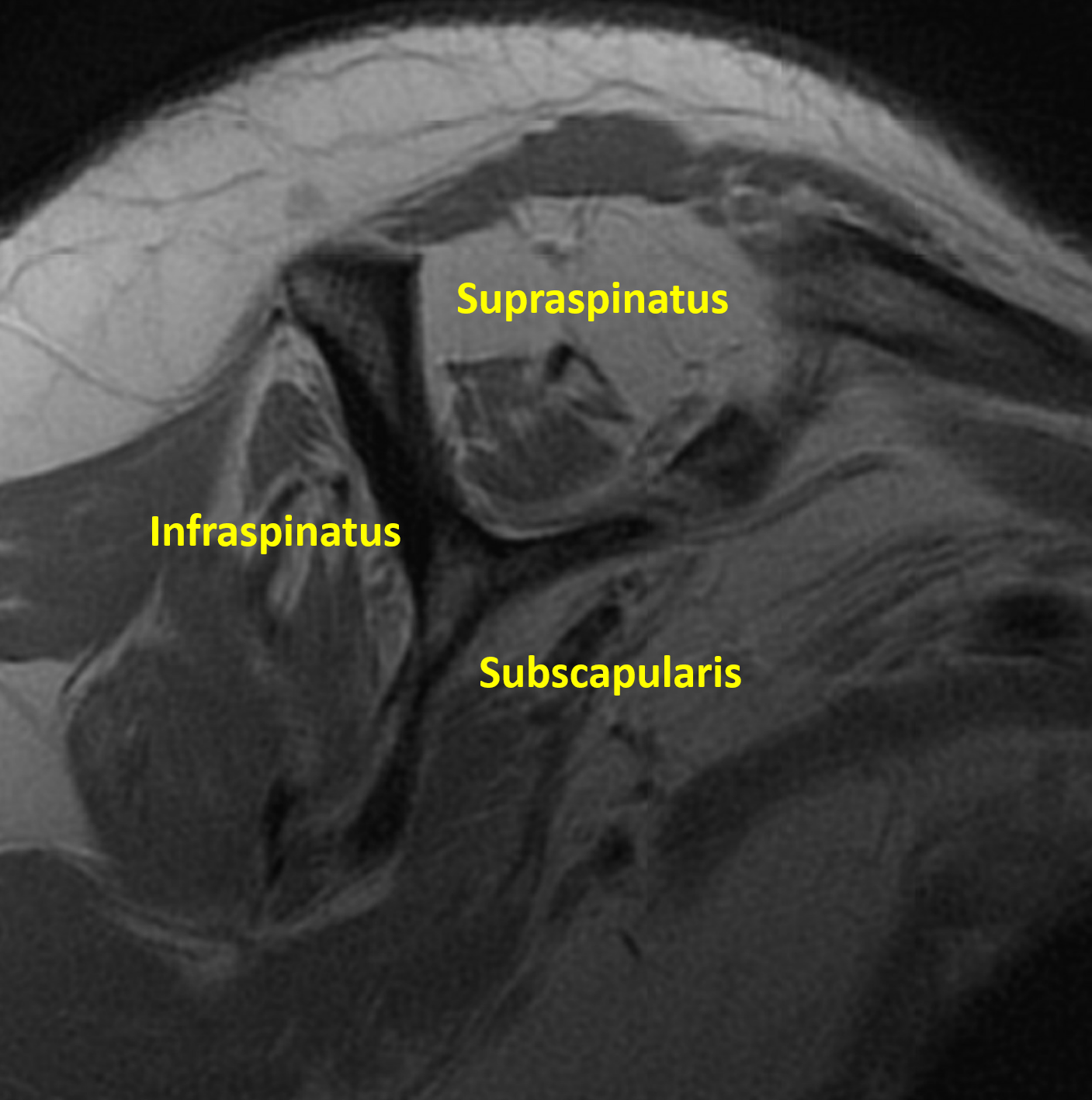
The Sagittal Plane depicts the shoulder as if viewing from the patient’s side. This plane allows a unique opportunity to see the rotator cuff muscles simultaneously and evaluate their quality. For instance, the image below depicts healthy rotator cuff muscles with appropriate size and homogenously dark color.
In contrast, this image depicts a sagittal MRI image that shows rotator cuff muscle atrophy and fatty infiltration. These features are critical to assess and have direct treatment implications for patients with rotator cuff injuries.
Disclaimer: The information provided on this website is intended for informational and educational purposes only. The opinions, views, and content presented here should not be construed as medical advice, diagnosis, or treatment. We welcome you to schedule a consultation with Dr. Brusalis for evaluation of your specific medical condition.


